INTRODUCTION TO MICROSCOPY AND MICROSCOPES
Microscopy is the technique involves the investigation of microorganisms or small objects that are not within the resolution range of the normal human eye. This can be done by using a special instrument called a microscope.
The microscope is an instrument that is used to produce an enlarged and well-defined image of objects, which are too small to be observed with the naked eyes. The most commonly used microscope in the laboratory is an Optical or light microscope.
The purpose of microscopy is to magnify the object so that one can easily observe the microbes, maximize the resolution to investigate the minute details precisely and to optimize the contrast between the structures, microbes, and background.
Nowadays the following types of microscopes are being employed for the study of microbes –
Check out the Normal Laboratory Values of Human body Parameters
⇒ Optical or Light microscope –

- The optical microscope contains a light source and a compound lens system, commonly used to examine the bacteria either in the living state or after fixing onto the slide and staining.
- It aids the examination of wet films or ‘hanging drops’ for the shape, arrangement, motility and rough idea of the size of the cells.
⇒ Phase-contrast microscope –
- It improves the contrast and makes evident the internal structures of cells which differ in thickness or refractive index.
- When light rays passed through an object and the surrounding medium, causes retardation of the light rays depending upon the differences in refractive indices, it produces ‘phase’ differences between the two types of rays.
- The phase differences produced are converted into differences in the intensity of light by a special optical system that produces light and dark contrast in the image.
⇒ Darkfield (dark ground) microscope –
- In this, the reflected light is used instead of the transmitted light used in the optical microscope, improves the contrast and produces better images of the internal structures of the cell.
- It consists of a darkfield condenser with a circular stop – fitted with a light microscope. This condenser lens system is arranged in such a way that no light reaches the eye unless reflected from the object.
- The object or bacterium appears self-luminous against a dark background.
Check out the List of Medical Doctors & Their Specialty
⇒ Fluorescent microscope –
- A fluorescent microscope is an optical microscope that uses fluorescence or phosphorescence to investigate the microbes.
- In this the specimens stained with fluorescent dyes like Auramine, Acridine orange etc. are exposed to the light of shorter wavelength – UV light or X-rays – results in the emission of longer wavelength visible light.
- This can be of two types –
- Fluorochrome – involves non-specific staining of bacteria with a fluorescent dye.
- Immunofluorescence – uses antibodies labeled with fluorescent dyes to specifically stain a particular bacterial cell.
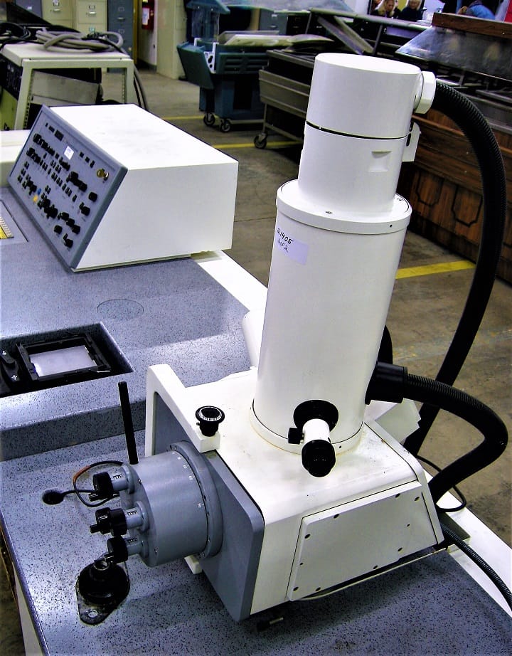
⇒ Electron microscope
- In this microscope, a beam of electrons is used instead of the beam of light used in the optical microscope.
- An electron beam is focused by circular electromagnets (magnetic condenser) – analogous to lenses of the light microscope.

- The wavelength of electrons used is 0.005nm as compared to visible light which is 500 nm
- Theoretically, the resolving power of the electron microscope should be 100000 times but practically, it is about 0.1 nm.
Check out the list of commonly used Medical Abbreviations

Hi, I’m the Founder and Developer of Paramedics World, a blog truly devoted to Paramedics. I am a Medical Lab Tech, a Web Developer and Bibliophiliac. My greatest hobby is to teach and motivate other peoples to do whatever they wanna do in life.