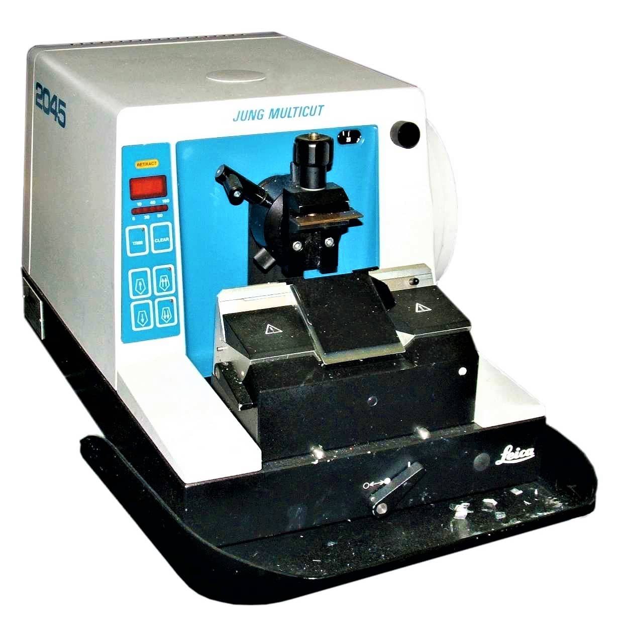
INTRODUCTION TO MICROTOMES
Microtome is an instrument with the help of which sections of tissues are cut and the process of cutting thin sections is known as Microtomy. The thickness of sections produced during microtomy may be between fractions of 50-100 nm, in ultramicrotomy, to several 100 microns. The common range is between 5-10m but both the maximum and minimum thickness is limited by the consistency of relation of the thickness of sections to the nature of tissues. These sections are stained using suitable staining techniques followed by observing them under the microscope.
TYPES OF MICROTOMES –
1.) Rotary microtome
The Rotary microtome is so-called because of a Rotary action of the handwheel responsible for the cutting moment. The block holder is mounted on a steel carriage, which makes up and down in groves this type of instrument is the most ideal for routine and research work it is excellent for cutting serial sections.
Parts of the rotary microtomes
- Block holder
- Knife clamp screw
- Knife clamps
- Block adjustment
- Thickness gauge
- The angle of tilt adjustment
- Operating handle

Here the feed mechanism is activated by turning a wheel on one side of the machine. The knife is fixed with its edge fixed upwards and the object is moved against the knife rising and falling vertically.
One rotation of the operating wheel produces a complete cycle downwards cutting stroke and an upward return stroke and activation of the advanced mechanism. It is often modified to cut ultrathin sections between 50Å – 200Å
The wheel may be electrically operated or manually. In the former case the hands may be made free for tissue maintenance, makes it available for incorporation in automated cryostats.
Advantages of the Rotary microtome
- Heavy and stable.
- Ideal for serial sections in large numbers.
- Paraffin-embedded tissues are cut by a rotary microtome.
- The knife holder is movable.
- The sections are cut are flat.
- It is useful for routine and research papers.
2.) Sliding or Base Sledge Microtome
This is a large heavy instrument with a fixed knife beneath which the object moves mounted on a heavy sliding base containing the feed mechanism used primarily for cutting the sections of cellulose nitrate embedded tissues with an obliquely set knife.
Parts of Base-sledge microtome
- Angular tilt adjustment
- Knife clamps
- Block holder
- Coarse feed adjustment
- Operating handle
- Thickness gauge
- Adjustment locking nut
- Block adjustment screw
- Split nut clasp
The blocks holder is mounted on a steel carriage which slides backward and forwards on groups against a fixed horizontal knife this microtome is heavy and very stable. The block is raised towards the knife at a predetermined thickness. This type of microtome is designed for cutting sections of very large blocks of tissues for example whole brain, this microtome has become popular for routine use.
Advantages of Base-sledge microtome
- It is useful for cutting extremely hard blocks and large sections.
- The microtome is heavy and stable.
- The knife used is sledge shaped which requires less honing.
3.) Cambridge rocking microtome
The instrument is so named because the arm has to move in a rocking motion while cutting the sections. The instrument was invented by Sir Horace Darwin in 1881 was developed by Cambridge company hence it is called the Cambridge rocking microtome. It is a simple machine in which the knife is held by means of microtome thread. The rocking microtome was designed primarily for cutting paraffin wax sections but in an emergency use frozen section by inserting a wooden block in which the tissue is frozen.
Parts of the rocking microtomes
- Knife holder
- Block holder or chuck
- Upper arm
- Screw
- Lever
- Pawl
- Ratchet wheel
- Mil head microtome screw
- Sleeve
- Lower Arm
- Scale
It cuts the sections between 1 to 20 microns. The knife is fixed with the edge, while the object is moved against this knife circularly, producing a sharply curved surface to the block with each stroke the tissue holder automatically moves vertically towards the life. Cutting stroke is Spring operated and is easy to handle. The microtome must be placed on a solid non-slippery surface to allow a better hold
Advantages of Cambridge rocking microtomes
- The cost of a knife and microtome is low.
- Celloidin embedded tissues can be sectioned easily.
4.) Freezing microtomes
This type has been designed for the production or preparation of frozen sections of fluid and non-fluid tissues usually without preliminary embedding. The object stage is connected to the cylinder of compressed carbon dioxide for the rapid cooling of the tissues and provisions are also made for the cooling of the knife.
Part of freezing type microtome
- Knife clamps
- Operating handle
- Thickness gauge
- Stage
- Stage valve
- Coarse adjustment
The movement of the knife takes place horizontally across the surface of the tissues. Ribbon sections cannot be prepared using this microtome. All freezing microtomes have the feature of employing a non-movable tissue block and cooling system.
Advantages of Freezing microtome
- It is used for sections required for Rapid diagnosis
- It cuts non-dehydrated fresh tissue in a frozen state.
- The method is useful for Rapid histopathological diagnosis during operation
- This type of microtome is also used when lipids, enzymes, and neurological structures are to be demonstrated.
Nowadays, the most commonly used type of microtome is a Rotary microtome which is easy to operate and ideal for routine use for diagnosis and research purposes.
WORKING PRINCIPLE OF ROTARY MICROTOME –
⇒ It is used for slicing paraffin tissue sections of uniform thickness.
⇒ This method is designed to cut 1-60 micron thick sections.
⇒ A knob on the device (typically at the backside) is used to modify the thickness of the sections.
⇒ A knife is constant inside the knife holder and clamped tightly.
⇒ The tissue block is drawn throughout the knife-edge and it is mechanically advanced. The top and bottom of the block have to be parallel and horizontal and as a minimum 1mm of paraffin has to be present in all aspects beyond the tissue.
⇒ The trimming of the edges of the block is usually completed with a single-sided razor blade and the block face is trimmed with the microtome knife.
⇒ The technician decides the type of section to be made in line with the nature of tissue and instructions received from the pathologist.
⇒ At some stage in section slicing, as the wheel of the microtome turns, sections are cut and slide on the knife. A ribbon of sections is produced.
⇒ The ribbon of sections is transferred to warm water inside the tissue floatation bath to put off any wrinkles present in the section.
⇒ The best quality section that is free from any scratches and cracks can be decided on from the tissue ribbons. The tissue ribbons are then taken on smooth glass slides with a respective identification number.
⇒ The slides are pulled from the water and the preferred sections are positioned flat on the surface of glass slides. The slides with the sections are positioned on a rack in a hot air oven to dry.

Hi, I’m the Founder and Developer of Paramedics World, a blog truly devoted to Paramedics. I am a Medical Lab Tech, a Web Developer and Bibliophiliac. My greatest hobby is to teach and motivate other peoples to do whatever they wanna do in life.
Uses of rotary microtme?
Rotary microtomes are commonly used to cut thin sections of paraffin-embedded tissues.
Procedures followed for staining for paraffin sections in microtomy
pls i need u to help me out i wrote a project base on this particular topic but i dont knw how to go about conclusion and recommendation maybe u can help me out
Kindly reach at info@paramedicsworld.com
Structural and functional difference between different types of microtome
Good attempt and informative for me