INTRODUCTION TO SUPERFICIAL MYCOSES
Superficial mycoses are the infections caused by fungi in the outermost layers of the skin, hair or nails.
Superficial mycosis is further classified as:-
- Surface infections
- Cutaneous infections
A.) SURFACE MYCOSES –
⇒ The fungi live exclusively on the dead layers of the skin, its appendages (Hair & Nail) and mucosa.
⇒ They have no contact with living tissue and elicit no inflammatory response.
⇒ For e.g. – Tinea versicolor, Tinea nigra and Tinea piedra.
Tinea versicolor (Pityriasis versicolor)
⇒ This is a chronic, usually asymptomatic involvement of the stratum corneum.
⇒ The disease is worldwide in distribution but is particularly prevalent in the tropical regions. It occurs mainly in young adults.
⇒ It is caused by the lipophilic, yeast-like fungus Malassezia furfur, formerly called as Pityrosporum orbiculare.
⇒ There is patchy discoloration or depigmentation of the skin occurs most commonly on the Chest, Abdomen, upper Limbs and Back.
⇒ Lab diagnosis of Pityriasis versicolor –
- Direct microscopy – Skin scraping is examined microscopically by KOH preparation, shows an abundance of yeast-like cells and short, branched
- Culture – the fungus can be grown on SDA media covered with a layer of olive oil in case of lipophilic fungus. Creamy colonies develop within 5-7 days at 37° Lactophenol cotton blue is used for stain.
Tinea nigra
⇒ It is characterized by black-brown macular lesions affecting the thick keratinized sites such as palms and soles.
⇒ This disease mainly occurs in the tropical regions.
⇒ It is caused by a fungus – Exophiala werneckii, formerly called as Cladosporium werneckii or Hortaea werneckii; and Exophiala castellanii.
⇒ Lab diagnosis of Tinea nigra –
- Skin scraping in 10% KOH mount shows brown, septate, branching Hyphae and budding cell.
- On SDA medium, moist, dark grey or black color colonies appear.
Tinea piedra
⇒ Tinea piedra is a fungal infection of hair.
⇒ The two varieties of Tinea piedra have been recognized as – Black piedra and White piedra.
⇒ The causative agent of Black piedra is Piedraia hortae, characterized by the presence of irregular black hard nodules on hair shaft of beard and scalp
⇒ The causative agent of White piedra is Trichosporon beigelii, characterized by white nodules on hair shaft of Axilla, Moustache, and Beard etc.
⇒ The nodules are composed of fungal elements cemented together on the hair.
⇒ Lab diagnosis of Tinea piedra –
- Hair mounted in 10% KOH solution shows the presence of nodules containing Asci having Ascospores.
B.) CUTANEOUS MYCOSES –
⇒ Cutaneous infections are mainly caused by a group of fungi called as Dermatophytes.
⇒ Dermatophytes –
- Dermatophytes are a group of fungi that live and infect only keratinized tissues of the body that are present superficially – Skin, Hair, and Nails.
- They metabolize keratinous tissues by unable to invade in living tissues.
- They cause Dermatophytoses commonly called as tinea or Ringworm.
⇒ Classification of Dermatophytes –
» Dermatophytes have been classified into 3 genera as follows –
GENUS INFECTED AREA
Trichophyton Hair, Skin, Nail
Microsporum Hair, Skin
Epidermophyton Skin, Nail
» Clinical classification of Dermatophytes is according to the site of lesions which is as follows –
DISEASE AFFECTED AREA
Tinea barbae (barber’s itch) Bearded areas of face and neck.
Tinea corporis (Tinea glabrosa) Ringworm of smooth or non-hairy skin.
Tinea imbricata Special type of Tinea corporis characterized
by Extensive concentric rings of
papulosquamous scaly patches.
Tinea capitis Ringworm of the scalp.
Tinea cruris (jock itch) Infection of groin and perineum.
Tinea pedis
(athlete’s foot)Ringworm of Foot
Tinea manuum Infection of hands
Tinea ungium Infection of Nails
» There are two variants of Tinea capitis – Favus and Kerion
- Favus is a chronic type of ringworm in which dense crust (scutula) develop in the hair follicles, leading to Alopecia and scarring.
- Kerion is characterized by severe boggy lesions with the marked inflammatory reaction that sometimes develops in scalp infection due to Dermatophytes.
» Dermatophytes and their common causative agents –
DISEASE COMMON CAUSATIVE AGENT
Tinea Capitis Any species of Microsporum and Trichophyton
Favus Trichophyton schoenleinii, Trichophyton violaceum, Microsporum gypseum.
Tinea barbae Trichophyton rubrum, Trichophyton mentagrophytes, Trichophyton verrucosum
Tinea imbricata Trichophyton concentricum
Tinea corporis Trichophyton rubrum
Tinea cruris Epidermophyton floccosum, Trichophyton rubrum.
Tinea pedis Trichophyton rubrum, Epidermophyton floccosum
Ectothrix hair infection Microsporum species,Trichophyton rubrum, Trichophyton mentagrophytes
Endothrix hair infection Trichophyton schoenleinii, Trichophyton tonsurans, Trichophyton violaceum
⇒ Clinical features of Dermatophytosis –
- Lesions in the skin – Circular, Dry, Erythematous, Scaly and Itchy.
- Lesions of hair – Kerion, Scarring, and Alopecia.
- Lesions of Nail – Deformed, Friable and discolored.
⇒ Laboratory diagnosis of Dermatophytes –
- Specimens: Skin, Hair and/or Nail, according to the site of the lesion. Scrapings are taken from the edges of ringworm lesions.
- Microscopy: a wet preparation of the specimen in 10% KOH solution may show the presence of Branching hyaline septate Hyphae, indicates the presence of Dermatophytes.
- Culture:
- For the culture, SDA or SDA media with antibiotics is used
- Culture plates are inoculated with the specimen and incubated at 25-30C for 2-3 weeks.
⇒ Identification of Dermatophytes –
» Identification of Dermatophytes in the laboratory is done by examining the macroscopic and the microscopic characteristics of the fungal colonies.
» Macroscopic examination – includes the rate of growth, texture, color on the obverse and reverse. Following are the characteristics of three genera –
- Trichophyton – colonies may be powdery, velvety or waxy, with pigmentation characteristics of different species
- Microsporum – colonies are cotton-like, velvety or powdery, with white to brown pigmentation.
- Epidermophyton – colonies are powdery and greenish yellow.

» Microscopic examination – Lactophenol cotton blue preparation from the colonies reveals microconidia, macroconidia or both as follows –
- Trichophyton – Microconidia are abundant, arranged in clusters along the Hyphae or borne on conidiophores. Macroconidia are scanty, elongated, with blunt ends and have distinctive shapes in different species which aids species identification.
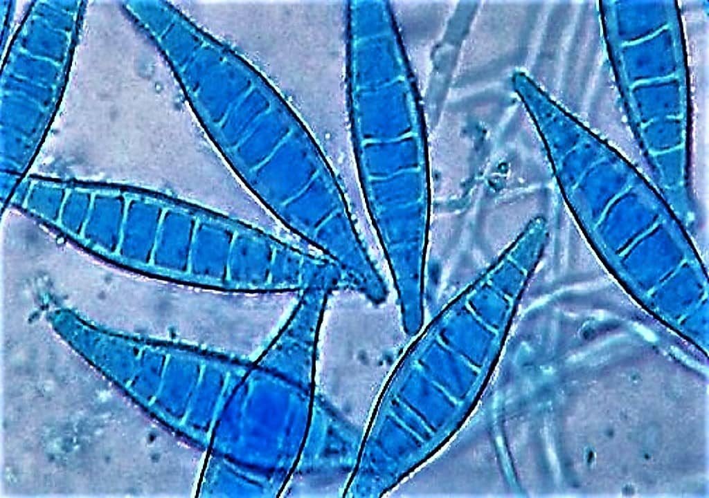
- Microsporum – microconidia are relatively scanty and not distinctive. Macroconidia, are large, Multicellular, spindle-shaped structures, borne singly on the ends of Hyphae.
- Epidermophyton – microconidia are absent. Macroconidia are Multicellular, pear-shaped & arranged in clusters.

Hi, I’m the Founder and Developer of Paramedics World, a blog truly devoted to Paramedics. I am a Medical Lab Tech, a Web Developer and Bibliophiliac. My greatest hobby is to teach and motivate other peoples to do whatever they wanna do in life.
this was helpful in my school project!